Subtotal $0.00
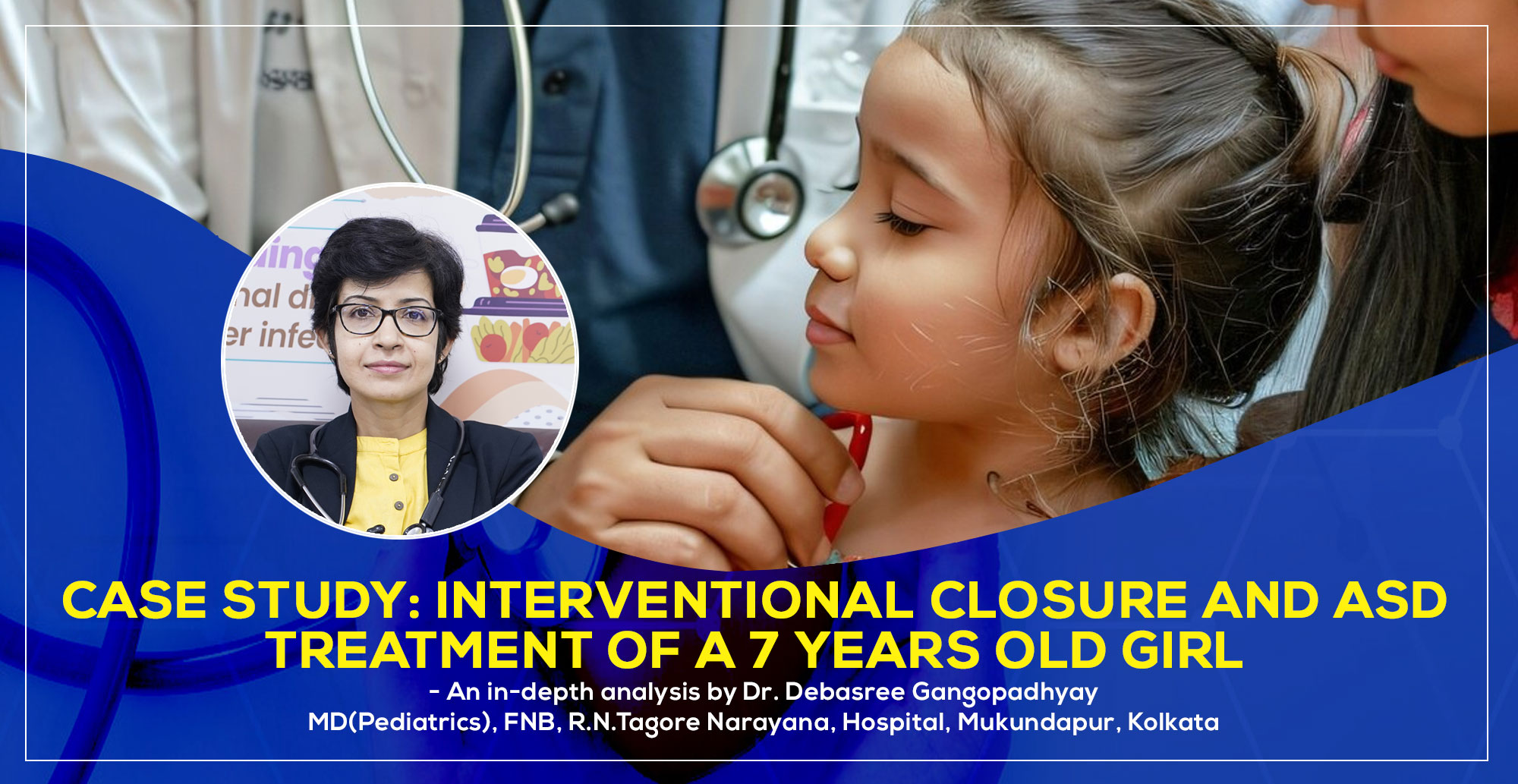
Case Study: Interventional Closure and ASD Treatment of a 7 years old girl
Introduction to Interventional Closure and ASD
Atrial Septal Defect (ASD) is a congenital heart defect characterized by a hole in the wall (septum) separating the heart’s two upper chambers. This condition leads toabnormal blood flow, straining the heart and lungs over time. This hole allows oxygen-rich blood from the left atrium to flow into the right atrium, mixing with oxygen-poor blood. This creates an extra volume of blood flowing to the lungs, causing increased strain on the heart and pulmonary system. An untreated ASD can lead to complications such as pulmonary hypertension, arrhythmias, heart failure, or stroke. The advancement of interventional cardiology has revolutionized the treatment of ASD, offering non-surgical solutions such as interventional closure. This case study explores the procedure and its impact, with a focus on the role of interventional cardiologists in Kolkata and congenital heart disease doctors in Kolkata.
This case study highlights the percutaneous device closure of a large ASD in a 7 year old patient, emphasizing diagnostic findings, procedural details, and follow-up outcomes.
What is an Atrial Septal Defect (ASD)?
ASD happens when there is a hole in the septum that separates the atria, which permits oxygen-rich blood from the left atrium to mix with oxygen-poor blood from the right atrium. The lungs and the right side of the heart may eventually become overworked as a result.
Common Causes of ASD
ASD typically results from genetic predispositions or developmental issues during pregnancy. Environmental factors may also contribute to its occurrence.
Symptoms of ASD
- Fatigue during exercise
- Shortness of breath
- Heart murmurs detected during routine check-ups
- Swelling in legs, feet, or abdomen
Case History: Clinical Presentation
Patient Initial Symptoms: A 7 year old girl was brought in after her parents noticed she frequently experienced fatigue and respiratory problems during physical activities, such as running and playing. The child’s growth and development were otherwise normal.
Physical Examination:
- Height and weight were appropriate for age.
- After a thorough clinical examination and imaging tests, including an echocardiogram, she was diagnosed with a large Atrial Septal Defect (ASD). The defect was enabling oxygen-rich blood from the left atrium to mix with oxygen-poor blood in the right atrium, causing increased workload on her heart and lungs.
- Cardiac examination revealed.
- A systolic murmur at the left upper sternal border and a fixed splitting of the second heart sound were detected by cardiac auscultation.
Diagnostic Evaluation
Echocardiography:
- Transthoracic echocardiography (TTE):Confirmed the existence of an 18 mm secundum ASD with left-to-right shunting and indications of right ventricular and atrial enlargement.
- Transesophageal echocardiography (TEE):Confirmed that there was sufficient rim tissue for device attachment byproviding thorough images of the defect’s size and edges.
- Electrocardiography (ECG):showed symptoms of volume overload, including right ventricular hypertrophy and right axis deviation.
- Chest X-ray:displayed enhanced pulmonary vascular outlines and massive heart, which are signs of left-to-right shunting.
Intervention
Pre-procedural Planning:
- Based on the defect features and lack of other structural heart abnormalities, the patient was considered for percutaneous device closure.
- Pre-procedure imaging confirmed suitability for interventional closure.
- After obtaining consent from her parents, the procedure was performed.
Procedure Details:
- Access:Under general anesthesia,the femoral vein was used to get percutaneous access while under general anesthesia.
- Imaging Guidance:To visualize the defect and direct device implantation, intraprocedural TEE wasused.
- Device Selection: A device was selected based on the defect’s size and surrounding tissue.
- Deployment:To ensure a safe implantation and the lack of residual shunting on post-deployment imaging, the device was enclosed in a delivery protective sheath.
- Complications:There were no immediate side effects from the surgery, including vascular injury, device embolization, or arrhythmias.
Post-Procedure Outcomes
Immediate Results:
- Post-procedural TEE confirmed complete closure of the defect and appropriate device positioning.
- The patient was transferred to the pediatric intensive care unit for monitoring after the procedure wassuccessfully performed using a septaloccluder device.
- The patient was discharged within 48 hours.
Outcome and Long-Term Prognosis
24-hour Monitoring: After 24 hours, the patient was hemodynamically stable and exhibited no evidence of residual shunting on the echocardiography report.
One-Month Follow-Up: After one month, clinical examination revealed that the murmur had resolved, and echocardiography revealed normal right heart size and function.
Six-Month Follow-Up: There was no sign of device migration, erosion, or arrhythmias, and the child continued to be asymptomatic.
Discussion
The treatment of secundum ASDs has been completely transformed by percutaneous device closure, which provides a minimally invasive option to surgical repair. In this case, the Amplatzer™ SeptalOccluder showed outstanding safety and effectiveness.
Advantages of Percutaneous Device Closure:
- Faster Recovery
- Reduced hospital stay
- Improved Quality of Life
- Avoidance of cardiopulmonary bypass and surgical scars
- Lower Risk of Complications
Limitations:
- Long-term monitoring is necessary to identify any late complications.
- In some cases, percutaneous closure may not achieve complete defect closure, necessitating further interventions.
- This treatment is not appropriate for ASDs with weak rims or other irregularities.
Conclusion
This case demonstrates the safety and effectiveness of percutaneous device closure in pediatric patients with secundum ASD.Early intervention can prevent long-term complications like pulmonary hypertension and arrhythmias, ensuring better quality of life and normal growth. Regular follow-up with echocardiography is critical to ensure long-term success. The expertise of the pediatric cardiologist in Kolkata and the advanced facilities available in Kolkata played a pivotal role in ensuring a successful outcome. Families dealing with similar conditions can find hope in the transformative impact of this modern procedure.
FAQs on Interventional Closure for ASD
What is the success rate of interventional closure for ASD?
The procedure boasts a success rate of over 95% in experienced hands.
Are there any risks involved in the procedure?
There are rare, potential risks, including device dislodgment or minor bleeding, which are manageable.
How soon can a child return to normal activities post-treatment?
Most children can return to their regular activities within a week.
Can ASD reoccur after treatment?
Once successfully closed, ASD is unlikely to reoccur.
How do I find the best pediatric cardiologist in Kolkata?
You should look for a specialist with experience in interventional cardiology and excellent patient reviews.

Dr. Debasree Gangopadhyay is a highly respected pediatric cardiologist based in Kolkata, India, specializing in the diagnosis and treatment of heart conditions in children. With a compassionate approach and a commitment to excellence, Dr. Gangopadhyay has made significant contributions to the field of pediatric cardiology. Her expertise includes managing congenital heart defects, arrhythmias, and other cardiovascular conditions in young patients. Dr. Gangopadhyay is dedicated to providing personalized care and staying updated with the latest advancements in pediatric cardiology. She is passionate about educating families on heart health and actively participates in research and community outreach programs.


Comments are closed